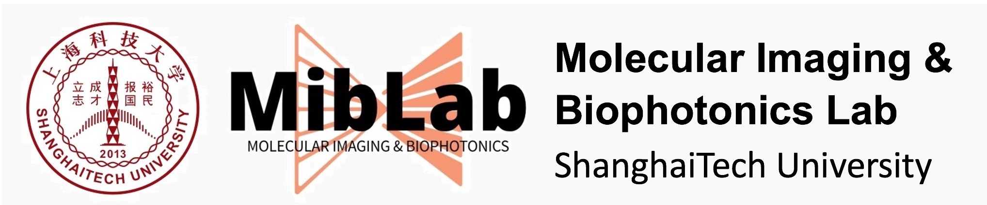
We are aiming at developing a novel optical imaging platform capable of visualizing multiple contrast parameters in biological tissues. The platform integrates the state-of-the-art hardware components such as wavelength-tunable pulse laser and time-of-flight cameras, as well as novel reconstruction algorithms and accurate calibration methods. Main application of such a platform includes small animal imaging and image-guided surgery.
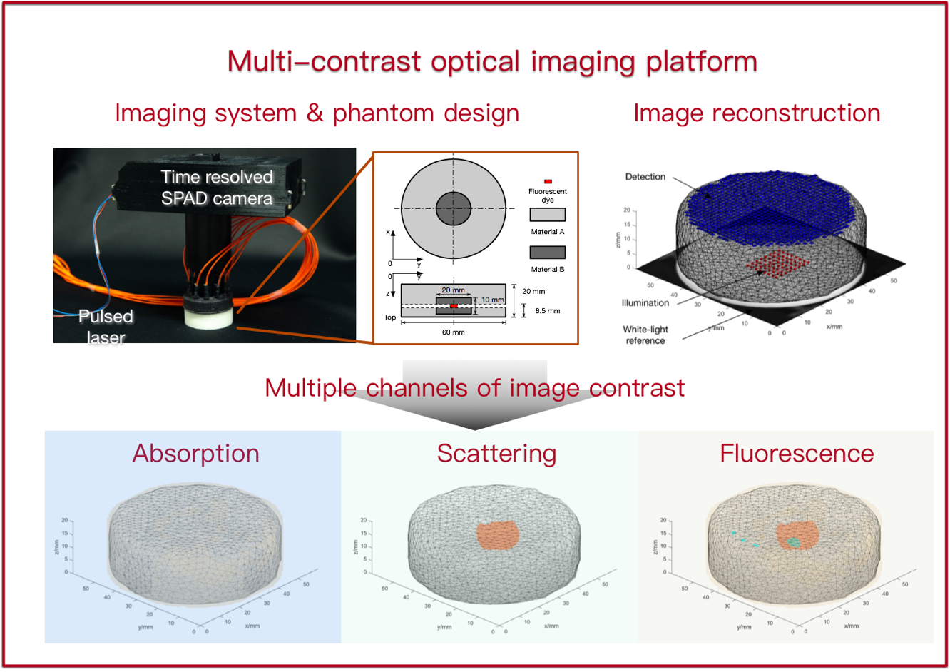
Multimodal imaging has emerged as a powerful method for biomedical research and clinical usage, as more comprehensive physiological information can be achieved compared with a standalone modality. In our lab, we combine optics and MRI due to several reasons. First, optical imaging, fluorescence imaging in particular, features high sensitivity and specificity which is complementary to MRI. Second, MRI can provides as an accurate anatomical reference that is useful for localizing the fluorescence signal. Third, both MRI and optical imaging can avoid ionizing radiation. Our team has previously developed the world’s first fluorescence tomography-MRI system and optoacoustic tomography-MRI system.
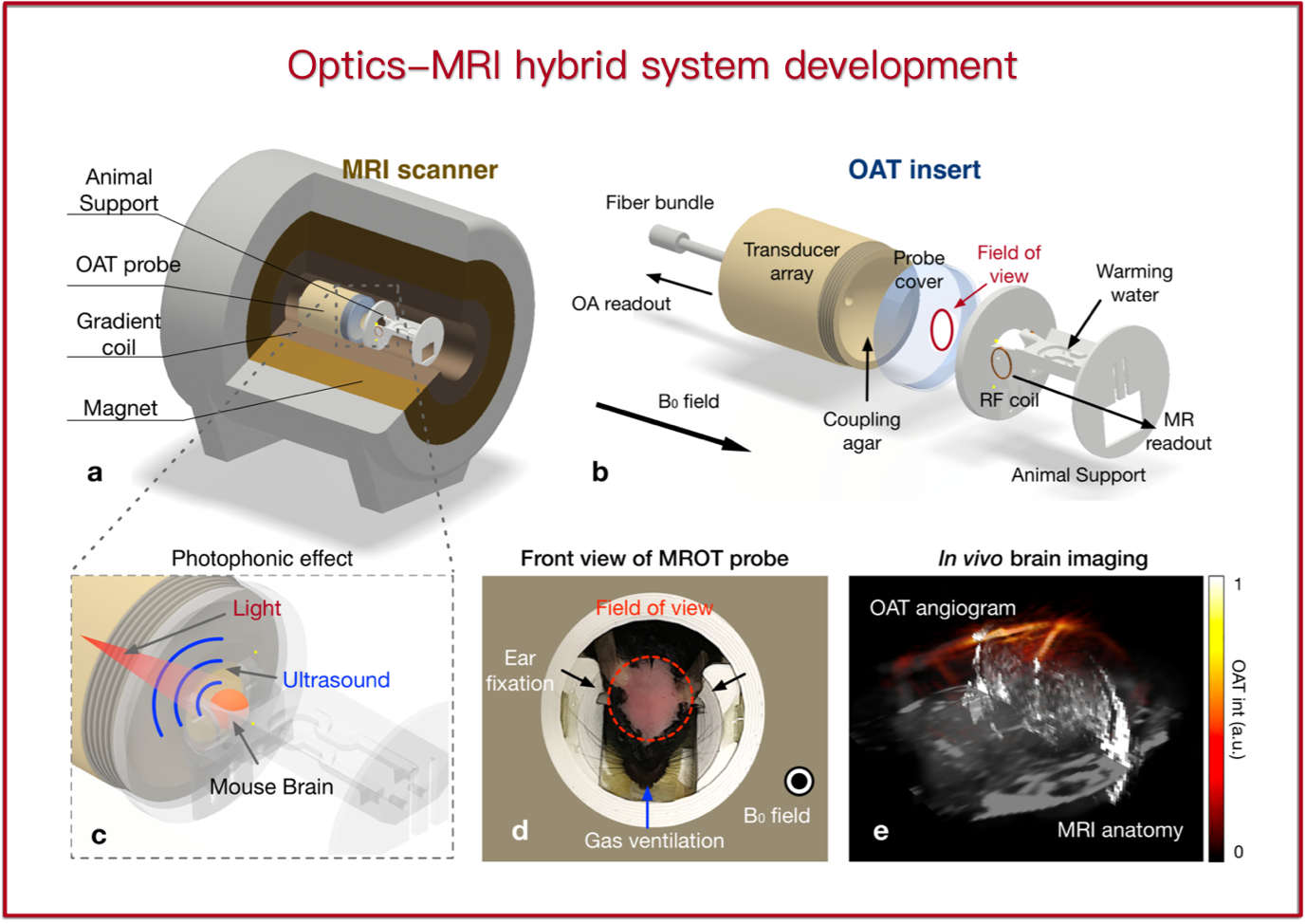
The conception of 3D reconstruction is commonly used in optical microscopy, e.g., in confocal microscopy and two-photon microscopy. However, visualizing objects deeply seated in tissue (> 1 mm) becomes challenging due to severe light scattering in tissue. Such a macroscopic reconstruction in scattering media is well known as a highly ill-posed inverse problem. We are seeking for fast, robust, 3D, high-resolution image reconstruction algorithms based on advanced computational techniques and novel machine learning methods.